Perspective Core Hub
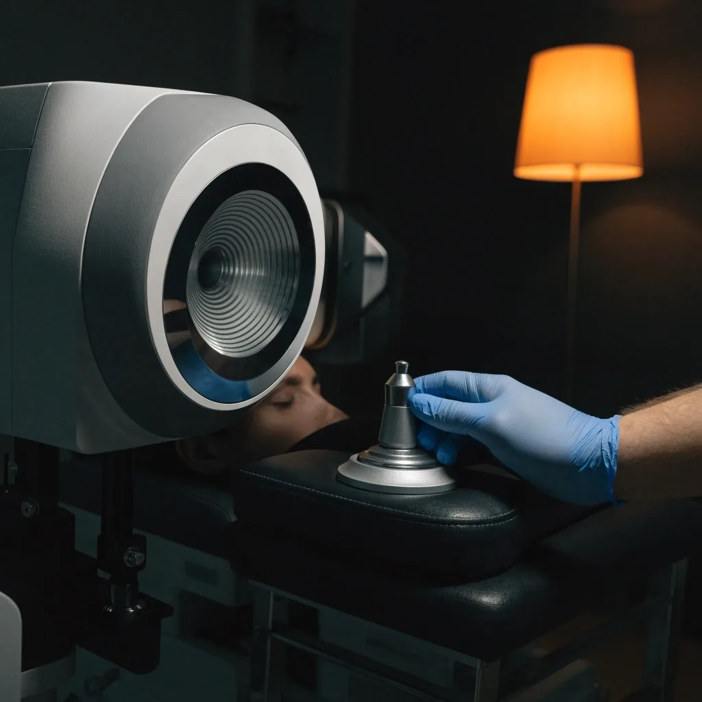
Ocular surface imaging platform in a darkened measurement room, adjusted by a gloved hand
Outcomes literature, read plainly

Outcomes & Predictability

Refractive predictability, corneal biomechanics and higher-order aberrations are the three measures outcome papers keep returning to. Here is what each one actually records, how it is reported, and what it cannot tell you.

What this page covers

Predictability is not a promise about your result. It describes how tightly a group of corrected eyes landed on their target correction, and how widely those results scattered around it. Biomechanics describe how the cornea holds its shape under that change. Higher-order aberrations are the optical irregularities beyond plain sphere and cylinder.

Outcome literature reports all three, but in different ways: predictability usually as a scatter or a percentage within a target band, biomechanics as measured response under load and across time, higher-order aberrations as wavefront terms. Reading them side by side tells you more than reading any one of them alone, because a technique can look tight on one measure and loose on another.

Below, each measure gets its own reading: what the number or term means, where the data comes from during a study, and which claims a careful reader should treat as provisional. This is educational material, not a personal prognosis.

How predictability is measured

Studies start by choosing a target: usually the correction each eye was programmed for. Before surgery, the measured refractive error is recorded. After surgery, the same measurement is repeated at defined visits. The difference between the achieved result and the target is the error the study tracks.

Plain-language reading of four commonly reported predictability measures
Reported measure What it records How a reader should treat it
Spherical equivalent error Distance from target in dioptres, treated as a signed number so over- and under-correction stay distinguishable The base measure. A tight average with a wide spread means the group did well on paper while individual eyes varied more
Within-target share How many treated eyes fell inside a stated band, such as within half a dioptre of target Only meaningful with the band named. A share quoted without its dioptre window tells you almost nothing
Stability over visits Change in refraction between follow-up points, usually months apart, rather than at a single endpoint The measure that answers whether the early result held. Single-visit reporting hides this entirely
Refraction at the corneal plane Where the correction is measured for the comparison, since spectacle-plane and corneal-plane values differ Check the convention. Mixing the two planes across studies makes groups look more different than they are

Reporting conventions and follow-up windows differ between studies. When you compare two papers, compare their definitions before you compare their numbers.

Corneal biomechanics: how the tissue answers

Removing or reshaping corneal tissue changes how the cornea carries load. Biomechanics is the branch of outcome research that asks how much stiffness was given up, and whether the remaining structure behaves predictably as pressure and healing act on it.

Investigators approach this from two directions. The first is direct measurement: instruments that record how the corneal surface responds to a controlled air pulse, or how it deforms under a known load. The second is indirect, reading shape change over time from topography and tomography and inferring what the response implies.

The practical takeaway for a patient is a boundary, not a score. Biomechanical measurements are most useful as an exclusion tool: they help a clinician decide which corneas should not be treated with a given technique at all. They are much less useful as a ranking of how good a result will be for an average eye.

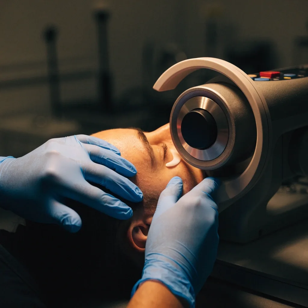
Gloved hands positioning a head on a corneal response measurement instrument
Biomechanical assessment is used mainly to decide whether a cornea should be treated with a given technique at all.

Higher-order aberrations in outcome reporting

Sphere and cylinder are the two corrections a spectacle prescription names. Everything else the optical system does is grouped as higher-order aberrations: coma, trefoil, spherical aberration and their neighbours. They are described as wavefront terms, which is why outcome papers sometimes refer to wavefront-guided or wavefront-optimised treatment rather than to plain correction.

Where the terms come from

Wavefront measurement passes light or a pattern through the eye and records how the returning front deviates from a perfect reference. The deviations are broken into named terms so the same measure can be compared between visits and between studies.

Why some terms matter more

A term that produces halo or starburst patterns around lights at night reads differently to a patient than a term that mainly affects fine contrast. Studies that report a single aggregate aberration number collapse that difference.

Where the optics meet the wound

Some change in aberration is expected after any tissue-removing procedure. Its size depends on the treatment profile, on centration, and on how the ocular surface behaves in the weeks after. A measured change is not automatically a problem.

Cross-study snag

Aberration values are strongly dependent on pupil size at measurement. A study taken at a larger pupil usually reports larger numbers. Comparing two papers without checking this understates or overstates the difference.

What improves over time

Some aberration change settles between the early and the later visits as the surface recovers. Reporting only the first postoperative measurement can make a temporary phase look permanent.

Treatment-design choice

Wavefront-guided and wavefront-optimised treatments differ in what they aim to conserve. Which one suits an eye depends on the pre-operative aberration profile and the tissue available, which is why the two are compared rather than ranked.


The three measures, side by side

Each measure answers a different question and only one of them is about your result as an individual. Reading them together is what outcome literature intends, even when a single paper emphasises just one.

Where you see a single strong value quoted online, check which of the three it is. A tight predictability number says nothing about tissue behaviour, and a favourable aberration profile says nothing about whether the correction held at month twelve.

Refractive predictability

Did the correction land where it was aimed?

Reported as distance from the intended correction and as the share of eyes inside a stated band. It is the measure most often quoted on its own, and the one most dependent on the band chosen.

Corneal biomechanics

Can the remaining tissue carry the new shape?

Read from response-to-load measurements and from shape change over time. Its clearest role in outcome work is exclusion, deciding who should not be scheduled for a given technique.

Higher-order aberrations

How clean is the optics beyond sphere and cylinder?

Measured as wavefront terms. Small changes after tissue removal are expected; the question papers ask is which terms changed, at what pupil size, and whether the change settled.

Measurements taken through a study

A published outcome is assembled from several points in time. Knowing where each number was captured explains most of the confusion when two papers appear to disagree. The frames below show the stages at which data is usually gathered in this kind of research.

Patient seated at a topographer during pre-operative refractive assessment
Before treatment, the measured error and the target correction are recorded separately.
Laser delivery cone above an eye on surgical drape during the treatment step
The treatment profile applied is the variable a study reports on, not the individual eye's outcome.
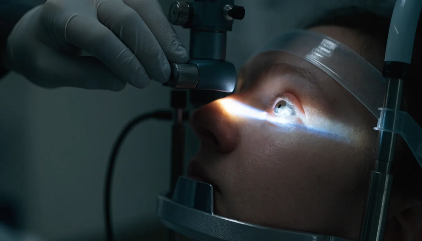
Surgeon examining an eye at a slit lamp during early follow-up
The first measured refraction is often a temporary reading, not the endpoint a study reports.
Patient reading an eye chart in a dimmed examination lane at a later follow-up visit
Stability is judged between later visits, which is where most disagreement between studies appears.
Measurement instrument calibration fixture under a focused work lamp between sessions
Instrument calibration is rarely discussed in outcome papers, yet it sets how comparable their measurements are.

Swipe or scroll the frames sideways on a narrow screen.

Questions readers ask about outcome numbers

These come up whenever someone tries to compare what they read online with what a clinic tells them in a consultation.

Why do two studies quote different predictability figures?

Usually because they defined their target band differently, or measured refraction at a different plane, or followed patients for a different number of months. The technique may be identical. Compare definitions before numbers.

Does a predictability figure predict my own result?

No, it does not. A group figure describes how a set of treated eyes behaved. Your eye has its own pre-operative measurements, its own healing response and its own ocular surface state. The value informs a conversation; it does not settle an outcome.

Why is corneal biomechanics discussed for exclusion rather than results?

Because the strongest evidence for its use is in identifying corneas that should not be treated with a given technique. Evidence that a biomechanical reading predicts how good a result will be for a typical eye is much weaker, so outcome papers do not lean on it that way.

Are higher-order aberrations always worse after treatment?

Some change is expected after any procedure that removes or reshapes tissue. What matters in the literature is which terms changed, whether the patient noticed a difference, and whether the change settled between the early and later visits.

How long does a study need to follow patients?

Long enough to include a stable phase rather than only the early healing weeks. Papers with twelve-month follow-up or longer answer more questions about stability; those with only the first month mostly describe the acute phase.

Where does ocular surface recovery fit into all this?

A surface that is still recovering changes refraction readings, so early measurements can look less stable than the eye's eventual state. That relationship between the surface and the readings is covered in its own chapter.

Perspective Core Hub · Jakarta

Where to go next with your questions

If a paper you have read raised a question about your own eyes, that belongs in a consultation rather than in a comparison of two studies. Bring the pre-operative measurements and any outcome figures you have seen; the useful conversation is about which measure applies to your case and which does not.

  • Jl. Cikini Raya No. 58, Menteng, Jakarta Pusat, DKI Jakarta 10330, Indonesia
  • +62 21 5189 4276
  • [email protected]
  • Monday to Friday 09:00–18:00 · Saturday 09:00–13:00